 Request Appointment
 Fine Needle Aspiration
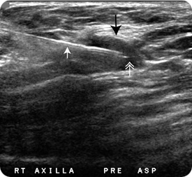
For fine needle aspiration, a small needle is used to aspirate, or suction, cells out of a lump in your breast or underneath your arm so that the cells can be evaluated under the microscope.
The procedure is safe, particularly when done with ultrasound guidance, but possible complications include:
- Infection
- Bleeding
- Pneumothorax (lung collapse) – an exceedingly rare complication, particularly when imaging guidance is used to sample the lesion
About the Procedure
During the procedure you usually lie on your back or slightly turned to one side with your arm placed comfortably under your head.
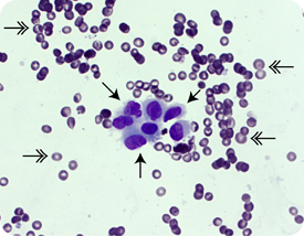
The skin is cleaned with betadine and alcohol and then numbed with topical anesthesia (1 percent lidocaine). A small needle is advanced into the lump under ultrasound guidance. As suction is applied, small movements are made with the needle in the lump to collect an adequate sample of cells. After the needle is pulled out of your breast, the aspirated material is smeared on a glass slide, fixed, stained and sent to pathology for review under the microscope by a cytopathologist.
At the completion of the procedure, firm pressure is held over the area sampled for approximately five minutes and an adhesive bandage is placed at the entry site on your skin. No restrictions or medications are indicated following the procedure
Your Results
Results of a fine needle aspiration are usually available within 24 hours. You will be asked to return to VCU Breast Imaging so that the radiologist can examine your breast and discuss the results with you.
It is important for the radiologist doing the procedure to correlate the imaging findings with what the pathologist describes on the samples. If the results are congruent and benign, it may be that nothing more needs to be done. If atypia or premalignant changes are described, a referral to a surgeon is made so that the area of concern can be completely removed surgically. If cancer is diagnosed, an MRI and consultation with a surgical oncologist are scheduled.
If there is insufficient material for the cytopathologist to make a diagnosis, or if the imaging and pathology findings are not congruent, a needle biopsy or consultation with a surgeon may be recommended.
“There is always a very professional, kind and caring staff here. Thank you for that. Keep it up.”
– Excerpt from a VCU Breast Imaging patient satisfaction survey.