Laser scanning microscopy provides for high-resolution, multi-channel (i.e. multi-color) three-dimensional imaging of cells and tissues. Its advantage over traditional widefield microscopy is that light scattering is reduced and out-of-focus “blur” is eliminated to yield “optical sections” of cells (or tissues) with higher image resolution. With confocal (i.e. single-photon) laser scanning microscopy, this is accomplished by illuminating the specimens with lasers (ultraviolet and visible light) that are scanned over the sample and imaging the fluorescence generated by the sample that passes through a pinhole aperture in the optical path. This system is ideal for high-resolution, three-D imaging of “fixed” (i.e. non-living) samples. However, there are significant limitations for imaging of living preparations:
- for live cell imaging, the short wavelength lasers are photo-toxic and can result in eventual cell death over relatively short imaging periods (i.e. after several minutes);
- the entire depth of the sample is illuminated by the laser resulting in photobeaching (i.e. fading) of the fluorescent tags over prolonged imaging periods;
- the short wavelengths of light used to illuminate the sample are more readily scattered by tissues, resulting in a limited imaging depth (100 - 200 µm).
Multi-photon (two-photon) laser scanning microscopy overcomes the limitations of confocal microscopy for prolonged live-cell and deep-tissue imaging. Multi-photon microscopy is based upon the fact that a fluorescent molecule may be excited (i.e. activated) by the simultaneous absorption of two photons that are approximately twice the wavelength of light used for conventional (single-photon) microscopy. Therefore, the microscope uses as its illumination source a pulsed, tunable, infrared (Ti:sapphire) laser.
There are several advantages for live-cell imaging:
- long wavelength light used for illumination is less photo-toxic than short wavelength light; therefore live samples may be imaged for prolonged periods (e.g. several hours) without any adverse affects.
- the photon density sufficient for 2-photon excitation occurs only at a narrow volume surrounding the imaging plane; therefore regions above and below the focal plane are not excited by the laser. This results in less photobleaching (i.e. greater stability of the fluorescent tags).
- since fluorescence occurs only in a narrow focal volume, a pinhole aperture is not required for confocal imaging, allowing for more of the light emitted by the sample to reach the detector.
- long wavelength light is less scattered than shorter wavelength light; therefore, samples may be imaged at significantly greater depths (200 - 800 µm).
The system is ideal for live-cell and live-tissue microscopy. This includes imaging of:
- cells growing in culture
- thick tissue slices,
- embryos and exposed tissues in situ.
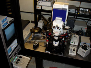
The facility houses a Zeiss LSM 510 META NLO multi-photon laser scanning microscope (fixed stage upright) with internal (descanned) detectors (including the META detector), two direct (non-descanned) detectors, and a transmitted light detector. The system has four lasers: a Spectra-Physics Mia-Tai broadband tunable Ti:sapphire laser (710-990 nm) for multi-photon imaging, as well as Argon (458, 476, 488, 514 nm), 561 diode (561 nm), and red HeNe (633 nm). Tuning of the Ti:sapphire laser is controlled directly via the LSM software. A number of high resolution and water immersion IR objective lenses are available for multi-photon imaging.
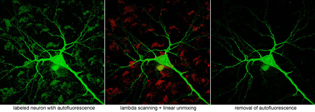
The META detector enables lambda scanning of fluorescence and emission "fingerprinting" as well as unmixing of signals from fluors with overlapping emission profiles.
For physiological imaging, a Luigs and Neumann 380 FM workstation and physiology recording equipment (stimulators, VP timing unit, preamp – A/D board, digitizer, multiclamp amplifier, temperature controller, in-line heater, peristaltic pump, etc.) are associated with the microscope. A Narishige PC-10puller is also available.