Variable pressure SEM allows for viewing conventionally prepared specimens under high vacuum conditions as well as viewing non-coated (non-conductive) and “wet” samples under low vacuum conditions. By introducing small amounts of air into the viewing chamber, molecules ionized by the electron beam neutralize the charge buildup on the specimen, thus permitting the sample to be viewed with the backscattered electron detector or the VPSE detector. This may help with the imaging of samples that are affected adversely by metal coating as well as samples that cannot be easily prepared by conventional methods for SEM. Hard biological samples may be viewed without any pretreatment (i.e. fixation, drying, coating) and soft “wet” samples (fixed or unfixed) may be viewed after introducing small amounts of water vapor into the column. The variable pressure system has an upper pressure limit of 750 Pa. The introduction of water vapor to the column insures that samples sensitive to desiccation are not dehydrated during imaging. Thus, wet biological samples may be imaged (with or without prefixation) without critical point drying, freeze-drying or metal coating.
Consultation, instruction, assistance, collaboration and service are provided. SEM services include sample preparation, fixation, critical point drying, sputter coating and SEM imaging.
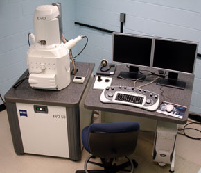
SEM equipment housed in the Microscopy Facility includes:
- Zeiss EVO 50 XVP scanning electron microscope (SEM) equipped with digital image acquisition, SE, VPSE & BSD detectors, extended variable pressure (up to 750 Pa), Deben coolstage and a water vapor introduction kit
- Tousimis autosamdri-814 Critical Point dryer
- EMS 550x sputter coater